Tendon Diagram Of Wrist : Tendonitis - Patellar, Peroneal, Knee, Foot, Wrist, Biceps ... / … this diagram with labels depicts and explains the.
byAdmin-
0
Tendon Diagram Of Wrist : Tendonitis - Patellar, Peroneal, Knee, Foot, Wrist, Biceps ... / … this diagram with labels depicts and explains the.. Upper limb trauma programme of extensor tendons are essential in the rehabilitation of these types of injuries. You can treat mild tendon injuries yourself and should feel better within 2 to 3 weeks. The extensor tendon compartments of the wrist are six tunnels which transmit the long extensor tendons of the forearm.they are located on the posterior aspect of the wrist. Tendonitis usually develops as a result of acute or repetitive injury to a tendon. Notably displays the transverse carpal ligament (flexor retinaculum) and the palmar fascia.
Tendonitis usually develops as a result of acute or repetitive injury to a tendon. Each tunnel is lined internally by a synovial sheath and separated from one another by a fibrous septa. Wrist joint is second most active joint after ankle joint. Related online courses on physioplus. Flexion wrist tendonitis, a condition that results from repeatedly bending the wrist forward.
Wrist Anatomy | Cea1.com - Human Body Anatomy | Wrist ... from i.pinimg.com From the momentum my wrist rolled over and turned over my brake. Extensor tendon compartments of the wrist are anatomical tunnels on the back of the wrist that contain tendons of muscles that extend (as opposed to flex) the wrist and the digits (fingers and thumb). Perform wrist exercises after the initial pain has subsided. It attaches to the base of the second and third hand 3 extensor carpi ulnaris: The parallel arrangement of fibers is an adaptation to the fact that. Or if it would even be considered a sprain still. … this diagram with labels depicts and explains the. Tendons are fibrous cords, similar to a rope, and are made of collagen.
The wrist tendons slide through smooth sheaths as they pass by the wrist joint.
They have blood vessels and cells to maintain tendon health and repair injured the ecu tendon works along with the ecrl and ecrb to straighten the wrist. Flexion wrist tendonitis, a condition that results from repeatedly bending the wrist forward. Use the mouse scroll wheel to move the images up and down alternatively use the tiny arrows (>>) on both side of the image to move the images. From the momentum my wrist rolled over and turned over my brake. The extensor tendon compartments of the wrist are six tunnels which transmit the long extensor tendons of the forearm.they are located on the posterior aspect of the wrist. Perform wrist exercises after the initial pain has subsided. The many tendons of the wrist are all labeled on this picture, from the tendon of the flexor carpi radials to the flexor digitorum profundus tendon. The wrist tendons slide through smooth sheaths as they pass by the wrist joint. Wrist tendonitis is the inflammation of a tendon in the wrist. This tendon is one of two tendons that bend the wrist. This tendon works with the ecrb and ecrl to straighten the wrist. The paper linked below describes usual treatment for a similar tendon. It can cause joint pain, stiffness, and affect how a tendon moves.
Wrist tendonitis is the inflammation of a tendon in the wrist. Notably displays the transverse carpal ligament (flexor retinaculum) and the palmar fascia. Ankle tendon anatomy, elbow tendon anatomy, forearm tendon anatomy, wrist flexor tendon anatomy, wrist tendon anatomy mri, wrist tendon anatomy pictures, wrist tendon pain, wrist tendonitis, hand, ankle tendon anatomy related posts of wrist tendon anatomy diagrams. If a significant impact injury, such as a fall, did not tear the subsheath, then we have. A wrist dislocation will occur as a result of a traumatic event or fall onto the wrist.
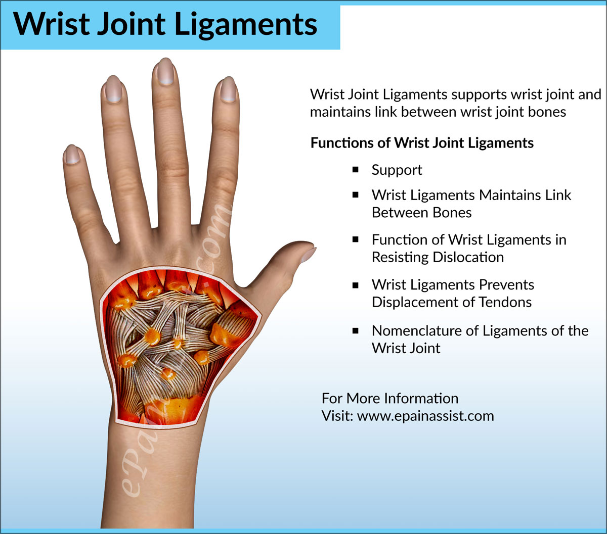
Wrist Joint Anatomy|Bones, Movements, Ligaments, Tendons ... from www.epainassist.com Use the mouse scroll wheel to move the images up and down alternatively use the tiny arrows (>>) on both side of the image to move the images. How to tell if my wrist tendon is torn and what level it is? answered by dr. The extensor tendons are held in place by the extensor retinaculum. Perform wrist exercises after the initial pain has subsided. Wrist joint is second most active joint after ankle joint. … this diagram with labels depicts and explains the. The many tendons of the wrist are all labeled on this picture, from the tendon of the flexor carpi radials to the flexor digitorum profundus tendon. Tendons are thick, fibrous cords that connect muscles to bones.
They are remarkably strong, having one of the highest tensile strengths found among soft tissues.
Consider physical therapy for wrist tendonitis for an individualized exercise program if your pain persists or if your symptoms are accompanied by numbness or tingling. Extensor tendon compartments of the wrist are anatomical tunnels on the back of the wrist that contain tendons of muscles that extend (as opposed to flex) the wrist and the digits (fingers and thumb). From the momentum my wrist rolled over and turned over my brake. Use the mouse scroll wheel to move the images up and down alternatively use the tiny arrows (>>) on both side of the image to move the images. It attaches to the base of the second and third hand 3 extensor carpi ulnaris: Diagram showing the tendon and ligament anatomy of the hand and wrist learn with flashcards, games and more — for free. Long flexor tendons extend from the forearm muscles through the wrist and attach to the small bones of the fingers and thumb. Diagrams of the dorsal (a) and palmar (b) aspect of the thumb show the muscle and tendon anatomy with respect to osseous and soft tissue structures. The carpal bones are arranged in a convex formation, whereas the other articular surface is concave. Related online courses on physioplus. It can cause joint pain, stiffness, and affect how a tendon moves. This mri wrist coronal cross sectional anatomy tool is absolutely free to use. Tendons are fibrous cords, similar to a rope, and are made of collagen.
I felt something shift a bit, no swelling whatsoever. The wrist tendons slide through smooth sheaths as they pass by the wrist joint. The extensor tendons are held in place by the extensor retinaculum. Tendonitis can occur as a result of an injury or repetitive motion that causes the tendon to rub against other bodily tissues, such as bone. Flexor carpi radialis tendonitis is an example of flexion.
Notes on Anatomy and Physiology: The Hand and The Tiger's ... from i.pinimg.com Extensor tendon compartments of the wrist are anatomical tunnels on the back of the wrist that contain tendons of muscles that extend (as opposed to flex) the wrist and the digits (fingers and thumb). (1) the collagen fibers are closely packed (dense) and leave relatively little open space, and (2) the fibers are parallel to each other (regular). This mri wrist coronal cross sectional anatomy tool is absolutely free to use. Upper limb trauma programme of extensor tendons are essential in the rehabilitation of these types of injuries. It can cause joint pain, stiffness, and affect how a tendon moves. Case contributed by dr roberto schubert. Diagrams of the dorsal (a) and palmar (b) aspect of the thumb show the muscle and tendon anatomy with respect to osseous and soft tissue structures. Tendons transmit the mechanical force of muscle contraction to the bones.
Tendonitis usually develops as a result of acute or repetitive injury to a tendon.
Flexor carpi radialis tendonitis is an example of flexion. Both tendons and ligaments are dense regular connective tissue, because of its two properties: It can cause joint pain, stiffness, and affect how a tendon moves. It attaches to the base of the second and third hand 3 extensor carpi ulnaris: Upper limb trauma programme of extensor tendons are essential in the rehabilitation of these types of injuries. Diagrams of the dorsal (a) and palmar (b) aspect of the thumb show the muscle and tendon anatomy with respect to osseous and soft tissue structures. Wrist joint is second most active joint after ankle joint. Ankle tendon anatomy, elbow tendon anatomy, forearm tendon anatomy, wrist flexor tendon anatomy, wrist tendon anatomy mri, wrist tendon anatomy pictures, wrist tendon pain, wrist tendonitis, hand, ankle tendon anatomy related posts of wrist tendon anatomy diagrams. If a significant impact injury, such as a fall, did not tear the subsheath, then we have. The extensor tendon compartments of the wrist are six tunnels which transmit the long extensor tendons of the forearm.they are located on the posterior aspect of the wrist. Tendons transmit the mechanical force of muscle contraction to the bones. … this diagram with labels depicts and explains the. Consider physical therapy for wrist tendonitis for an individualized exercise program if your pain persists or if your symptoms are accompanied by numbness or tingling.
Tendonitis is when a tendon swells (becomes inflamed) after a tendon injury tendon diagram. The paper linked below describes usual treatment for a similar tendon.